animal cells pictures labeled
Cheek Epithelial Cells: How to Prepare a Wet Mount Microscope Slide. 17 Images about Cheek Epithelial Cells: How to Prepare a Wet Mount Microscope Slide : Plant and Animal Cells - Labeled Graphics, Animal Cell Structure And Function Masteringbiology / Cell Cycle and also Plant and Animal Cells - Labeled Graphics.
Cheek Epithelial Cells: How To Prepare A Wet Mount Microscope Slide
 www.youtube.com
microscope cells epithelial wet mount cheek cell slide prepare animal specimen biology step eukaryotic slides inside
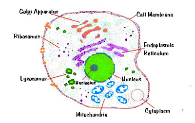
Biology Journal: ANIMAL CELL (SECONDARY SCHOOL STYLE)
 jamielee16106biologyjournal.blogspot.com
biology
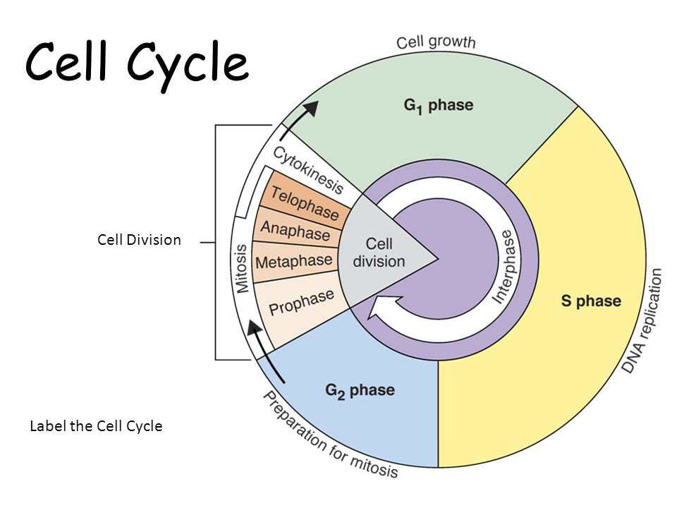
Phases Of Cell Cycle - Online Biology Notes
 www.onlinebiologynotes.com
cell cycle division phases mitosis phase biology g2 dna notes
Biology: The Animal Cell: Task
 questgarden.com
cell task animal biology cells version
Cells In Tissues, Organs And Systems | Cells As The Basic Units Of Life
 www.siyavula.com
cell animal drawing simple grade science natural cells plant basic mitochondria typical nucleus drawings structures dna sciences structure draw units
Animal Cell Diagram - Labeled | Science! | Pinterest | Search And
 www.pinterest.com
cell animal labeled diagram label exchange smarttech science
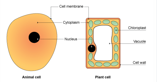
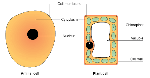
Plant And Animal Cells - Labeled Graphics
 www.biologycorner.com
animal cells labeled plant cell
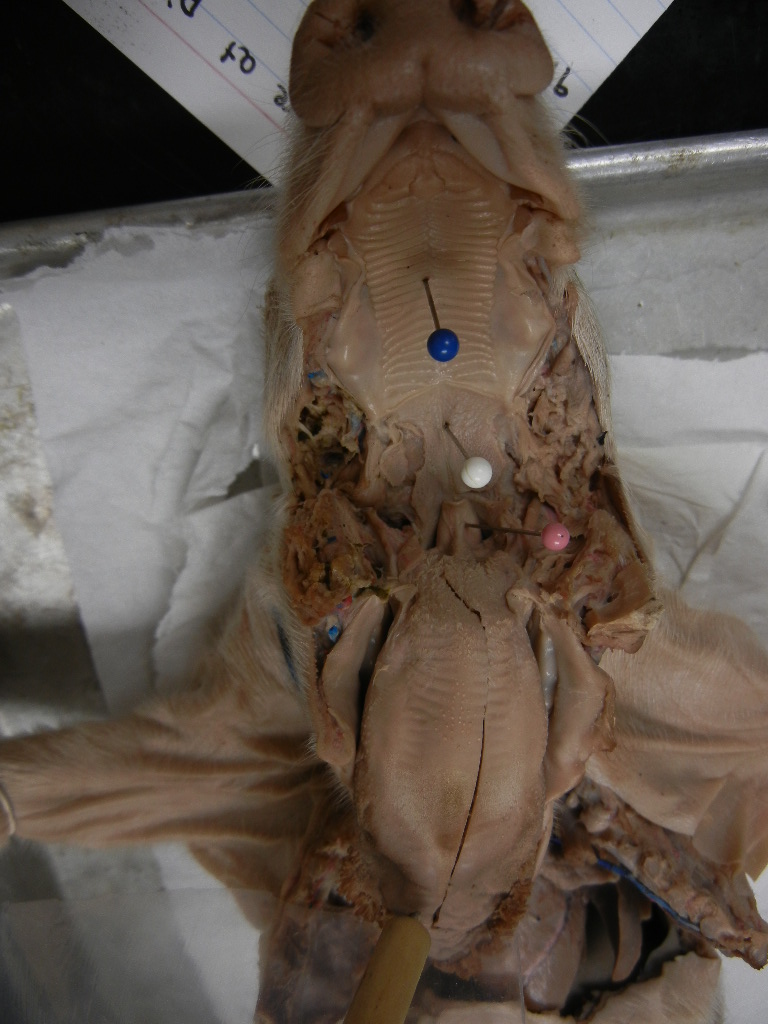
Fetal Pig Dissection Images
 www.biologycorner.com
pig fetal dissection labeled esophagus palate biology corner trachea
Animal Cell Structure And Function Masteringbiology / Cell Cycle
 robertpurkhisere02773.blogspot.com
purkhiser
Science In Year 5: Animal Cells And Plant Cells
 teacherdeliascienceyear5.blogspot.com
animal plant cells cell science simple ks3 bitesize diagram bbc parts gcse functions organelles differences nucleus between membrane cytoplasm unit
Quia - Animal Cell
cell animal quia
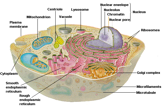
Cell Organelles- Structure And Functions With Labeled Diagram | Cell
 www.pinterest.ph
cell organelles diagram functions structure animal labeled human list function organelle plant cells
Cells: Animal Cells And Plant Cells
 sukhjeevankaur.blogspot.com
animal plant cells cell science simple ks3 bitesize diagram bbc parts gcse functions organelles differences nucleus between membrane cytoplasm unit
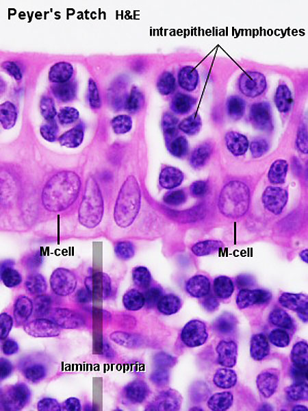
File:Peyer's Patch 02.jpg - Embryology
 embryology.med.unsw.edu.au
peyer lymphatic embryology freeworkshop unsw
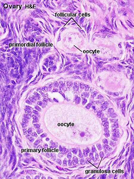
File:Ovary Follicle 01.jpg - Embryology
 embryology.med.unsw.edu.au
ovary follicle reproductive histology embryology ovarian albuginea tunica gland embryo primordial ovaries physiology oogenesis granulosa jejunum cortical oocytes stages menstrual
File:Vagina Histology 02.jpg - Embryology
 embryology.med.unsw.edu.au
histology embryology unsw
The Cell | Biochem Rocks
 kimberlybiochemist.wordpress.com
cells
Ovary follicle reproductive histology embryology ovarian albuginea tunica gland embryo primordial ovaries physiology oogenesis granulosa jejunum cortical oocytes stages menstrual. File:peyer's patch 02.jpg. Animal cell diagram